Elevated PSA
What a raised PSA result means, and how it is investigated — with MRI first and, when needed, a targeted biopsy.
An elevated PSA result is a common reason for referral to urology. It is understandably worrying, but a raised PSA is not a diagnosis. PSA can rise for many reasons, many of which are not cancer. This page explains what PSA is, why it can be raised, how the result is interpreted, and the steps that may follow — including MRI and, for some men, a prostate biopsy.
What is PSA?
Prostate-specific antigen (PSA) is a protein made by the prostate gland. Its normal role is to help keep semen fluid. A small amount passes into the bloodstream, where it can be measured with a simple blood test.
PSA is made by both normal and cancerous prostate tissue, so the test cannot tell the two apart on its own. Many men with a raised PSA do not have prostate cancer, and a normal PSA does not completely rule it out. For this reason, PSA is interpreted alongside your age, prostate size, family history and, increasingly, an MRI scan.
Why can PSA be raised?
The causes fall broadly into two groups: benign (non-cancerous) conditions and prostate cancer.
| Benign (non-cancerous) | Cancer |
|---|---|
| Benign prostatic enlargement (BPE) — the prostate naturally grows with age Inflammation or infection of the prostate (prostatitis) or a urinary tract infection Recent ejaculation Recent vigorous exercise, particularly cycling A recent catheter, cystoscopy or prostate biopsy Difficulty emptying the bladder (urinary retention) | Prostate cancer |
Temporary causes. Because several of these causes are short-lived, a raised result is often repeated before further tests. Ejaculation and vigorous exercise are usually avoided for 48 hours before a PSA test, and testing is postponed for at least 4 weeks after a urinary infection.
Medicines that lower PSA. Some medicines for an enlarged prostate — finasteride and dutasteride — lower the PSA reading by roughly half. If you take one of these, please tell your doctor, as your result needs to be interpreted with this in mind.
What counts as an elevated PSA?
PSA naturally rises with age, as the prostate grows. For this reason, the National Institute for Health and Care Excellence (NICE) in the UK uses age-specific thresholds.
| Age | PSA threshold (ng/mL) |
|---|---|
| Under 40 | Clinical judgement |
| 40–49 | More than 2.5 |
| 50–59 | More than 3.5 |
| 60–69 | More than 4.5 |
| 70–79 | More than 6.5 |
| Over 79 | Clinical judgement |
These thresholds are a guide to when further assessment is needed, and there is some variation between recommendations. A single result is always considered together with the trend over time, family history and general health.
PSA density
A larger prostate naturally makes more PSA. PSA density adjusts for this by dividing the PSA level by the volume of the prostate, which is measured on an MRI scan.
For example, a PSA of 6 in a 60 mL prostate gives a PSA density of 0.10, whereas the same PSA of 6 in a 30 mL prostate gives a PSA density of 0.20. The second result is more concerning, because more PSA is being produced for the size of the gland.
As a reference point, a PSA density of 0.15 or above may lead us to consider a biopsy when the MRI shows an uncertain area (PI-RADS 3). This is a reference value rather than an absolute rule — the decision always takes your overall picture into account.
The role of MRI
A multiparametric MRI scan of the prostate is now recommended for men with a raised PSA, before a prostate biopsy is considered. MRI can show areas of the prostate that look suspicious, help decide whether a biopsy is needed at all, and act as a map to guide the biopsy needle to the right place.
Two large UK-led studies changed practice. The PROMIS study found that MRI detected 93% of clinically significant cancers, compared with 48% for a standard transrectal biopsy, and suggested that around a quarter of men could safely avoid an immediate biopsy. The PRECISION trial then showed that an MRI-first approach, with targeted biopsy only where the scan was suspicious, found clinically significant cancer in 38% of men compared with 26% using standard biopsy — while diagnosing fewer insignificant cancers. In PRECISION, 28% of men in the MRI group avoided a biopsy.
MRI is very helpful, but it is not perfect. It can occasionally miss cancer, and it can show areas that turn out to be benign (false positive).
The PI-RADS scoring system
Radiologists report prostate MRI scans using a standard five-point scale called PI-RADS (Prostate Imaging Reporting and Data System). The score describes how likely it is that a clinically significant cancer is present.
| Score | Likelihood of clinically significant cancer |
|---|---|
| 1 | Very low — highly unlikely |
| 2 | Low — unlikely |
| 3 | Intermediate — uncertain |
| 4 | High — likely |
| 5 | Very high — highly likely |
Do I need a biopsy?
A biopsy is the only way to confirm prostate cancer, but not every man with a raised PSA needs one. The decision brings together your PSA, PSA density, MRI score, family history, age and general health, and is made together with you.
Deciding whether to have a PSA test in the first place →
- PI-RADS 1–2: a biopsy can often be avoided after discussion, with PSA monitoring instead. A biopsy may still be advised if other factors are concerning, such as a high PSA density, a rising PSA or a strong family history.
- PI-RADS 3: the result is uncertain, so the decision is individual. PSA density is particularly helpful here, with 0.15 used as a reference point.
- PI-RADS 4–5: a biopsy is usually recommended.
A biopsy is not usually recommended on the basis of a PSA result alone.
MRI fusion (targeted) biopsy
In an MRI fusion biopsy, your MRI images are combined (“fused”) with a live ultrasound image of the prostate during the procedure. This allows the biopsy needle to be guided to the specific area seen on the scan.
Two types of sample are usually taken. Targeted cores are taken from the suspicious area(s) identified on MRI. Systematic cores are also taken from the rest of the prostate, to reduce the chance of missing a cancer outside the target area. The samples are then examined under a microscope by a pathologist.
Transperineal or transrectal biopsy?
There are two routes for taking prostate biopsies, and both are offered in this practice.
Transperineal biopsy. The needle passes through the skin between the scrotum and the back passage (the perineum), rather than through the rectum. Because the bowel is avoided, the risk of infection is very low and preventive antibiotics are not required. This route gives good access to the front (anterior) part of the prostate, which can be harder to reach through the rectum. Transperineal biopsy is commonly performed under sedation for patient comfort.
Transrectal (TRUS) biopsy. The needle passes through the wall of the rectum under ultrasound guidance. It is performed under local anaesthetic and is generally well tolerated. To lower the risk of infection, a rectal swab is taken beforehand so that antibiotics can be targeted to the bacteria present. With this approach, the risk of infection is around 1%.
What the evidence shows. The UK TRANSLATE trial compared the two routes in 1,126 men who had an MRI before their first biopsy. Clinically significant cancer (Gleason 3+4 and above) was found in 60% of men who had a transperineal biopsy, compared with 54% with a transrectal biopsy. This difference is thought to reflect better sampling of the front and tip of the prostate with the transperineal route. Infection requiring hospital admission occurred in fewer than 1% of men after transperineal biopsy and 2% after transrectal biopsy.
The US PREVENT trial compared transperineal biopsy without antibiotics against transrectal biopsy with rectal swab-guided, targeted antibiotics. There were no infections after transperineal biopsy, and the infection rate after transrectal biopsy was relatively low at 1.4%. Detection of clinically significant cancer was similar with both routes.
| Transperineal | Transrectal (TRUS) | |
|---|---|---|
| Needle route | Through the skin of the perineum | Through the wall of the rectum |
| Anaesthetic (this practice) | Generally sedation, for comfort | Local anaesthetic |
| Preventive antibiotics | Not required | Targeted, guided by a rectal swab |
| Infection risk | Very low | Around 1% with targeted antibiotics |
| Sampling the front of the prostate | Good access | Can be harder to reach |
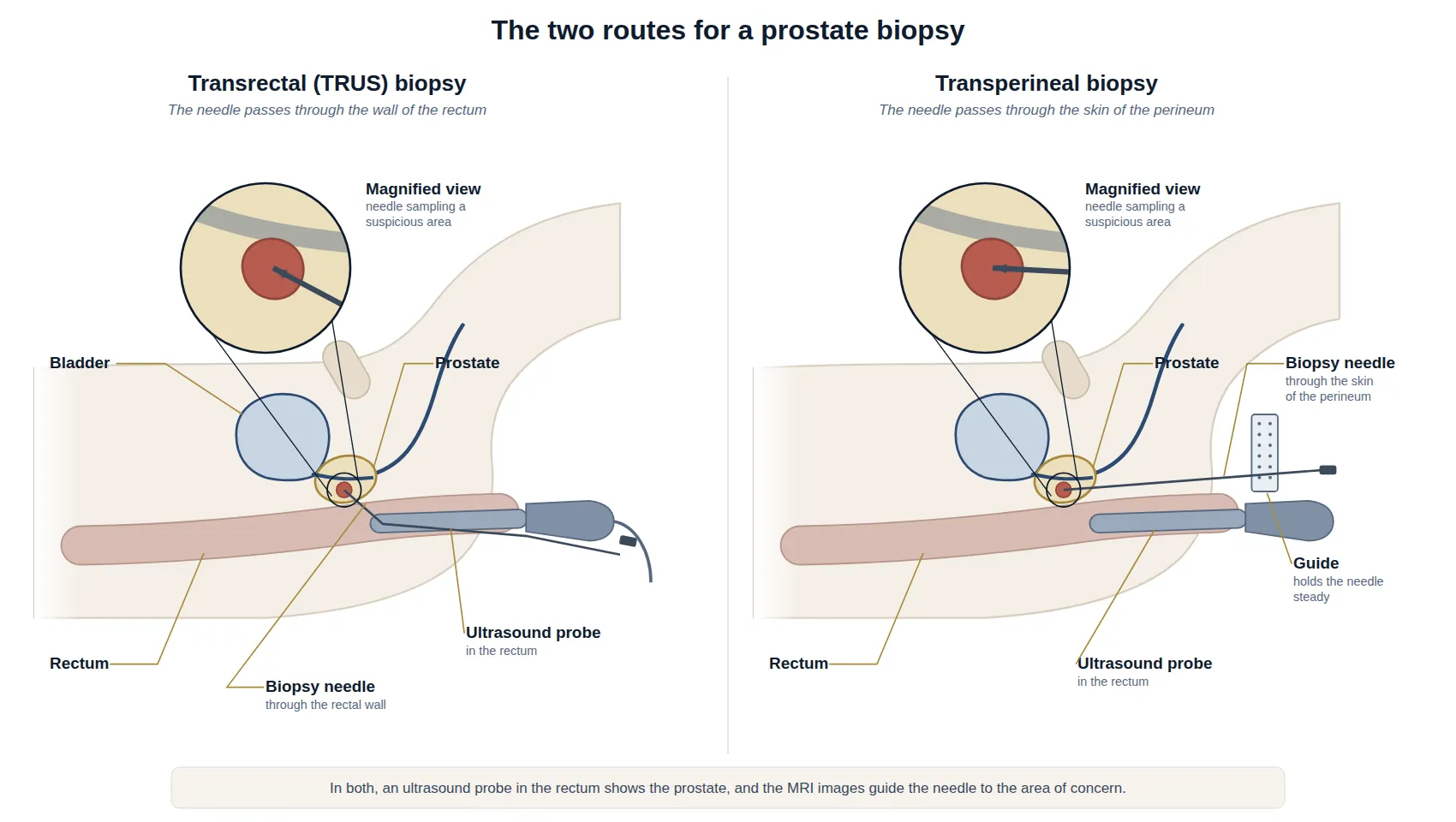
With either route, it is common to notice blood in the urine, semen or stools for a few days to weeks afterwards. A small number of men have temporary difficulty passing urine. The most suitable route for you will be discussed at your consultation.
Have a question about this condition?
Dr Tan consults at SJMC, Kuala Lumpur.