Understanding Bladder Cancer
What bladder cancer is, the single most important distinction your team will make, and what the first tests and operations involve.
Being told you may have bladder cancer raises many questions. This article explains what bladder cancer is, the single most important distinction your team will make, and what the first tests and operations involve. Understanding the vocabulary early makes the conversations that follow considerably easier.
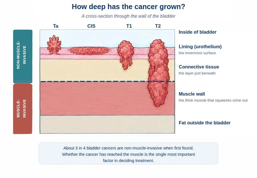
The bladder is a hollow, muscular bag that stores urine. Its inner surface is lined by a specialised layer of cells called the urothelium, and almost all bladder cancers begin in this lining. Underneath the lining sits a thin layer of connective tissue, and beneath that lies the thick muscle wall that squeezes urine out when you pass water.
Risk factors
Smoking is the single biggest risk factor. Long-term exposure to certain industrial chemicals — particularly in the dye, rubber and paint industries — also raises the risk. Bladder cancer becomes more common with age and is more frequent in men, although it is often diagnosed later in women, because blood in the urine is more readily attributed to other causes.
The most important question: has it reached the muscle?
Almost every treatment decision follows from a single question — whether the cancer has grown into the muscle wall of the bladder.
Non-muscle-invasive bladder cancer sits in the lining, or in the connective tissue just beneath it, but has not reached the muscle. Around three in four bladder cancers are non-muscle-invasive when first diagnosed. It is generally treated by removing the tumour completely — both to treat it and to obtain tissue for analysis — and, where appropriate, by giving medicine directly into the bladder. It tends to come back, so long-term monitoring is essential, but it does not usually threaten life.
Muscle-invasive bladder cancer has grown into the muscle wall. It is less common but considerably more serious, because it carries a real risk of spreading elsewhere. It requires major treatment — either removing the bladder, or treating it with radiotherapy, usually after a course of intravenous systemic therapy.
| Stage | What it means | Group |
|---|---|---|
| Ta | A growth on the surface of the lining | Non-muscle-invasive |
| CIS | A flat, high-grade patch within the lining | Non-muscle-invasive |
| T1 | Grown into the connective tissue beneath the lining | Non-muscle-invasive |
| T2 | Grown into the muscle wall | Muscle-invasive |
| T3 | Grown through the muscle into surrounding fat | Muscle-invasive |
| T4 | Grown into a nearby organ | Muscle-invasive |
Grade: how the cells behave
Alongside stage, the pathologist reports the grade, which describes how abnormal the cells look under the microscope. Low-grade cells resemble normal cells, grow slowly and rarely invade. High-grade cells look markedly abnormal and behave more aggressively. A small low-grade tumour and a small high-grade tumour are managed quite differently, so grade matters as much as stage.
You may also hear the term urothelial carcinoma, the medical name for the usual type that arises from the lining. A minority are of other types — described as histological subtypes — which can behave differently and may be treated more aggressively.
TURBT: the first operation
If a tumour is seen during a telescopic bladder examination in the clinic (a cystoscopy), the next step is almost always a transurethral resection of bladder tumour (TURBT). Despite the long name, it is a relatively low-risk procedure, and no cuts are made in your skin.
It is usually performed under general anaesthesia. A telescope is passed along the urethra into the bladder, and the tumour is shaved away and removed in pieces, with the base sealed to stop bleeding. Care is taken to include some underlying muscle in the sample — while minimising the risk of perforation — because this is what allows the pathologist to say whether the muscle is involved.
The operation therefore serves two purposes: it removes the visible cancer, and it provides the tissue that determines the stage and grade. You can read more about TURBT here.
What recovery is like
Most people go home the same day or after one night. A catheter — a soft tube draining the bladder — is often left in for one to two nights. Expect some blood in the urine and a burning sensation when passing water for one to two weeks; drinking plenty of fluid helps. Avoid heavy lifting for about two weeks. Contact your team if you develop a fever, cannot pass urine, or are passing large clots.
Other tests you may have
If the tumour is muscle-invasive or high-grade, you will need scans to check whether the cancer has spread — usually a CT scan of the chest, abdomen and pelvis.
A urine sample may be sent for cytology, looking for abnormal cells shed into the urine. It is helpful when positive, particularly for high-grade disease and CIS, but a normal result does not exclude cancer.
Can a urine test replace the camera test?
This is among the questions patients ask most often, and it is one our group has worked on for many years. At present, the available urinary tests do not have the diagnostic accuracy needed to replace cystoscopy.
We also asked patients themselves what they thought. In interviews and surveys, we found that most people would accept a urine test in place of cystoscopy — but only if it were nearly as accurate, and many valued the reassurance of a surgeon looking directly at the bladder. For now, cystoscopy remains the standard.
Getting the results
The tissue takes approximately one week to be examined. You will then be given a plan based on the stage, the grade, and your general health.
Sometimes a second TURBT is advised a few weeks later, particularly for some high-grade or T1 tumours. This is not a sign that something went wrong — it is done to be certain nothing was left behind and that the depth has been correctly assessed, because getting the stage right at the outset is crucial to recommending the correct treatment.
Living with the diagnosis
The period between the operation and the results is often the hardest part, and finding it difficult is entirely normal. In a study of how patients experience being diagnosed with bladder cancer, the waiting and the uncertainty were described as harder than the procedures themselves.
Practical steps help. Bring someone with you to the results appointment, as two people remember more than one. Write your questions down beforehand, since it is easy to forget them in the room. Ask for information in writing. And if smoking has been part of your life, stopping now genuinely improves your outlook — your team can arrange support.
Have a question about this condition?
Dr Tan consults at SJMC, Kuala Lumpur.
Bladder cancer is most often found after blood in the urine.