Renal Mass
What a renal mass is, why most are found by chance, and how scans, size and sometimes a biopsy guide what happens next.
Being told that a scan has shown a “mass” in your kidney is understandably worrying. A renal mass simply means an area in the kidney that looks different from the normal kidney tissue. Some are fluid-filled cysts, some are benign (non-cancerous) growths, and some are kidney cancers (most often renal cell cancer), often found at a small, early stage. This page explains how renal masses are found, the different types, how the risk of cancer is assessed, and when a biopsy may help.
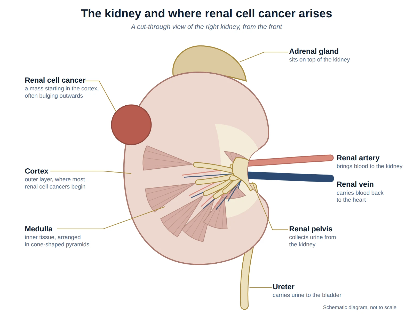
The kidneys are two bean-shaped organs, roughly the size of a fist, that sit high at the back of the abdomen. They filter waste from the blood to make urine, and also help control blood pressure and the production of red blood cells.
How renal masses are found
Most renal masses are now found by chance (incidentally), on an ultrasound, CT or MRI scan done for another reason — for example, to investigate abdominal pain, back pain or a problem with another organ. As scans have become more widely used, more kidney masses are being found, and they are usually found while still small and confined to the kidney.
Less commonly, a renal mass is found because of symptoms. These can include blood in the urine, a persistent ache in the side or back, or a lump — but these tend to be later signs, possibly associated with larger renal masses and occasionally with cancer that has spread (metastatic disease). Most small masses cause no symptoms at all.
Types of renal mass
Cystic masses. Simple kidney cysts — small, fluid-filled sacs — are very common, particularly with age. They are benign and usually need no treatment or follow-up. Cysts with more complex features are graded by radiologists using the Bosniak classification, which describes what the cyst looks like on a CT or MRI scan and how likely it is to be a cancer. The higher the category, the greater the chance of cancer.
| Category | What the scan shows | Chance of cancer | Usual approach |
|---|---|---|---|
| I | A simple cyst — thin, smooth wall and clear fluid, with no internal divisions, calcium or solid areas | Benign | No follow-up needed |
| II | A minimally complex cyst — a few thin internal divisions (septa), fine calcium, or dense fluid that does not take up contrast | Benign | No follow-up needed |
| IIF | Slightly more complex — a smooth, minimally thickened wall or divisions, or many thin divisions, but no solid areas. The “F” stands for follow-up | Low — under 1% if the cyst stays stable on follow-up scans | Follow-up scans over several years to confirm it is stable |
| III | Thickened or irregular walls or divisions that take up contrast | About 50% | Treatment or, in selected cases, careful monitoring — discussed individually |
| IV | A cyst containing a solid (nodular) area that takes up contrast | About 90% | Usually managed as a renal cell cancer |
Chance-of-cancer figures are pooled estimates from a systematic review of more than 3,000 complex kidney cysts (Schoots et al., 2017). Around 1 in 8 IIF cysts change to a higher category during follow-up, and these carry a high chance of cancer — which is why follow-up scans matter.
Solid masses. Most solid renal masses turn out to be renal cell cancer (kidney cancer), but not all. A proportion are benign growths:
- Angiomyolipoma (AML): a benign growth containing fat, blood vessels and muscle. It can often be recognised confidently on a CT or MRI scan by the fat it contains, so a biopsy is usually not needed.
- Oncocytoma: a benign growth that can look very similar to renal cell cancer on a scan, and is often only confirmed with a biopsy or after it has been removed.
Renal cell cancers themselves also vary. The main types — clear cell, papillary and chromophobe — are described on the Understanding Renal Cell Cancer page.
Assessing a renal mass
A renal mass is usually assessed with a CT or MRI scan using contrast (dye). This shows the size and position of the mass, whether it takes up the contrast (a feature of solid growths), whether it contains fat, and whether there is any sign of spread. If cancer is suspected, a scan of the chest may also be arranged. Blood tests check your kidney function, which is important when planning any treatment.
For many masses, the scan appearance together with the size gives a good indication of what the mass is likely to be, and helps decide whether monitoring, treatment or a biopsy is the most appropriate next step.
Size and the risk of cancer
Size is one of the most useful clues. Smaller solid renal masses are more likely to be benign, and when they are cancers, they are more likely to be slow-growing. As a mass gets larger, the chance that it is a cancer — and that the cancer is more aggressive — increases.
| Size of mass | Benign (not cancer) | Cancer |
|---|---|---|
| Under 4 cm | About 20% | About 80% |
| Over 4 cm up to 7 cm | About 10% | About 90% |
| 7 cm or larger | About 6% | About 94% |
Source: a large series of 2,935 solid renal tumours removed at surgery (Frank et al., 2003). Figures describe groups of patients and cannot predict an individual result.
The role of a renal mass biopsy
A renal mass biopsy takes small samples of the mass using a thin needle, guided by an ultrasound or CT scan, usually under local anaesthetic, sometimes with sedation. The samples are examined under a microscope to find out whether the mass is benign or cancer, and if cancer, what type.
A biopsy is not always needed. When the scan appearance is characteristic, or when the result would not change the plan — for example, in a fit patient where removing the mass (often with a kidney-sparing partial nephrectomy) would be recommended either way — treatment can proceed without a biopsy.
A biopsy can be helpful when the result may change the plan, for example:
- before choosing active surveillance (monitoring) for a small mass
- before ablation (destroying the mass with heat or freezing)
- when the scan appearance is uncertain, or another condition such as lymphoma, infection, or spread from a different cancer is possible
- before starting drug treatment for renal cell cancer that has spread
How accurate is it? A large review of 57 studies including more than 5,000 patients found that a renal mass biopsy gave a diagnosis in around 92% of cases. When core biopsy samples gave a diagnosis, they correctly identified cancer in 99% of cases (sensitivity) and correctly identified benign masses in close to 100% of cases (specificity). Biopsies are less reliable at predicting the grade of a cancer. If a biopsy does not give a clear answer, it may be repeated.
How safe is it? Serious complications are uncommon; minor bleeding is the most common. Biopsies are taken using a coaxial technique — a thin outer guiding needle is placed once, and the samples are taken through it — which protects the path of the needle. The risk of cancer cells spreading along the needle track (seeding) is very rare.
What happens next
The plan depends on the type and size of the mass, your kidney function, your general health and your preferences. The main options include:
- Monitoring (active surveillance): regular scans for small masses, particularly in older patients or those with other health problems.
- Partial nephrectomy: removing the mass while preserving the rest of the kidney — preferred wherever safe and feasible.
- Radical nephrectomy: removing the whole kidney, for larger or more complex masses.
- Ablation: destroying selected small masses with heat or freezing.
- Stereotactic radiotherapy (SBRT): an option where surgery or ablation is not possible.
These options are explained in more detail on the Treating Localised Renal Cell Cancer page.
Have a question about this condition?
Dr Tan consults at SJMC, Kuala Lumpur.