Understanding Renal Cell Cancer
What renal cell cancer is, how it is found and staged, and the language your team will use when discussing treatment.
A guide for patients: what renal cell cancer — the most common type of kidney cancer — is, how it is found and staged, and the language your team will use when discussing treatment. Understanding these terms early makes the decisions ahead far easier.
The kidneys are two bean-shaped organs, roughly the size of a fist, that sit high at the back of the abdomen. They filter waste from the blood to make urine, and also help control blood pressure and the production of red blood cells. Most kidney cancers begin in the tiny filtering tubes of the kidney and are called renal cell cancer (or renal cell carcinoma). This is different from cancer that begins in the lining of the drainage system, which is called upper tract urothelial cancer and is covered separately.
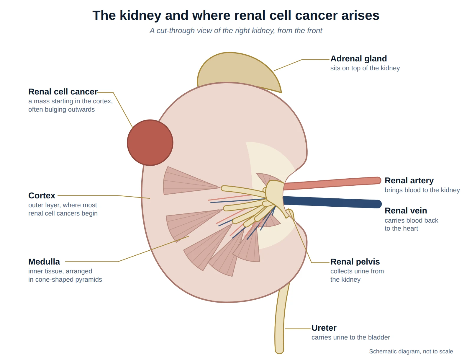
Types of renal cell cancer
Renal cell cancer is not a single disease. The pathologist identifies the type from the tumour, and the type can influence how it behaves and how it is treated.
| Type | How common | In brief |
|---|---|---|
| Clear cell | About 7 in 10 | The commonest type; the one most systemic treatments were designed around |
| Papillary | About 1 in 10 | A distinct type that can be less responsive to some drug treatments |
| Chromophobe | About 1 in 20 | Usually less aggressive; tends to have a good outlook |
| Other / rare | The remainder | A range of less common types, some managed at specialist centres |
Who it affects and why
Renal cell cancer becomes more common with age and is more frequent in men. Known risk factors include smoking, obesity, high blood pressure, and long-term kidney disease or dialysis. A small proportion of renal cell cancers run in families; if you are young at diagnosis, have cancer in both kidneys or several tumours, or have a strong family history, your team may suggest a genetic assessment.
How it is usually found
Many renal cell cancers today are found by chance, when a scan is done for another reason. When symptoms do occur, they may include blood in the urine, a persistent ache in the side or back, or a lump — but these are late signs, and most small cancers cause none. Because incidental discovery is now common, many renal cell cancers are found while still small and confined to the kidney.
Making the diagnosis
A renal cell cancer is usually identified on a CT or MRI scan, which shows its size, position, and whether it has spread. A biopsy is not always needed before treatment, because the scan appearance is often characteristic — although it can be useful in selected cases, for example before ablation, before drug treatment, or when the diagnosis is uncertain.
Grade and stage
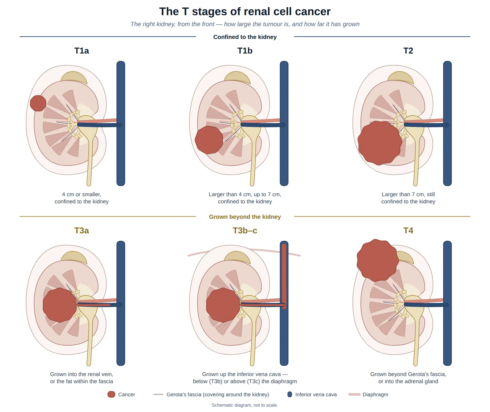
Two pieces of information shape the plan. The grade describes how aggressive the cells look under the microscope, reported on a scale from 1 to 4 (the WHO/ISUP grade): low grade (1–2) tends to behave less aggressively, and high grade (3–4) more aggressively. Grade is one factor among several — alongside the type and the stage. The stage describes how large the tumour is and how far it has spread, and is described using the TNM system.
| Category | What it describes | In brief |
|---|---|---|
| T — Tumour | Size of the tumour and how far it has grown | T1a: 4 cm or less, confined T1b: 4–7 cm, confined T2: over 7 cm, confined T3a: into the renal vein or the fat around the kidney T3b–c: into the large vein (inferior vena cava) T4: grown beyond the kidney’s covering, or into a nearby organ |
| N — Nodes | Whether it has reached nearby lymph glands | N0: no nodes involved N1: nearby nodes involved |
| M — Metastasis | Whether it has spread to distant sites | M0: no distant spread M1: distant spread (e.g. lung, bone, liver) |
| Stage | Roughly what it means | 5-year survival* |
|---|---|---|
| Stage 1 | Small, confined to the kidney (T1) | around 90% |
| Stage 2 | Larger, still confined (T2) | around 75% |
| Stage 3 | Into a vein or the surrounding fat, or nearby lymph nodes (T3, or N1) | around 75% |
| Stage 4 | Spread to distant parts of the body (M1) | around 15% |
*These are general figures for large groups of people (Cancer Research UK; patients diagnosed in England, 2016–2020) and cannot predict any individual’s outcome. Survival also depends on the cancer type, grade, and your general health, and newer drug treatments are improving outcomes — particularly for advanced disease.
Have a question about this condition?
Dr Tan consults at SJMC, Kuala Lumpur.