Focal Therapy
for Prostate Cancer
Treating only the part of the prostate that contains the cancer, rather than removing or irradiating the whole gland — with the aim of controlling the cancer while preserving urinary and sexual function.
What focal therapy is
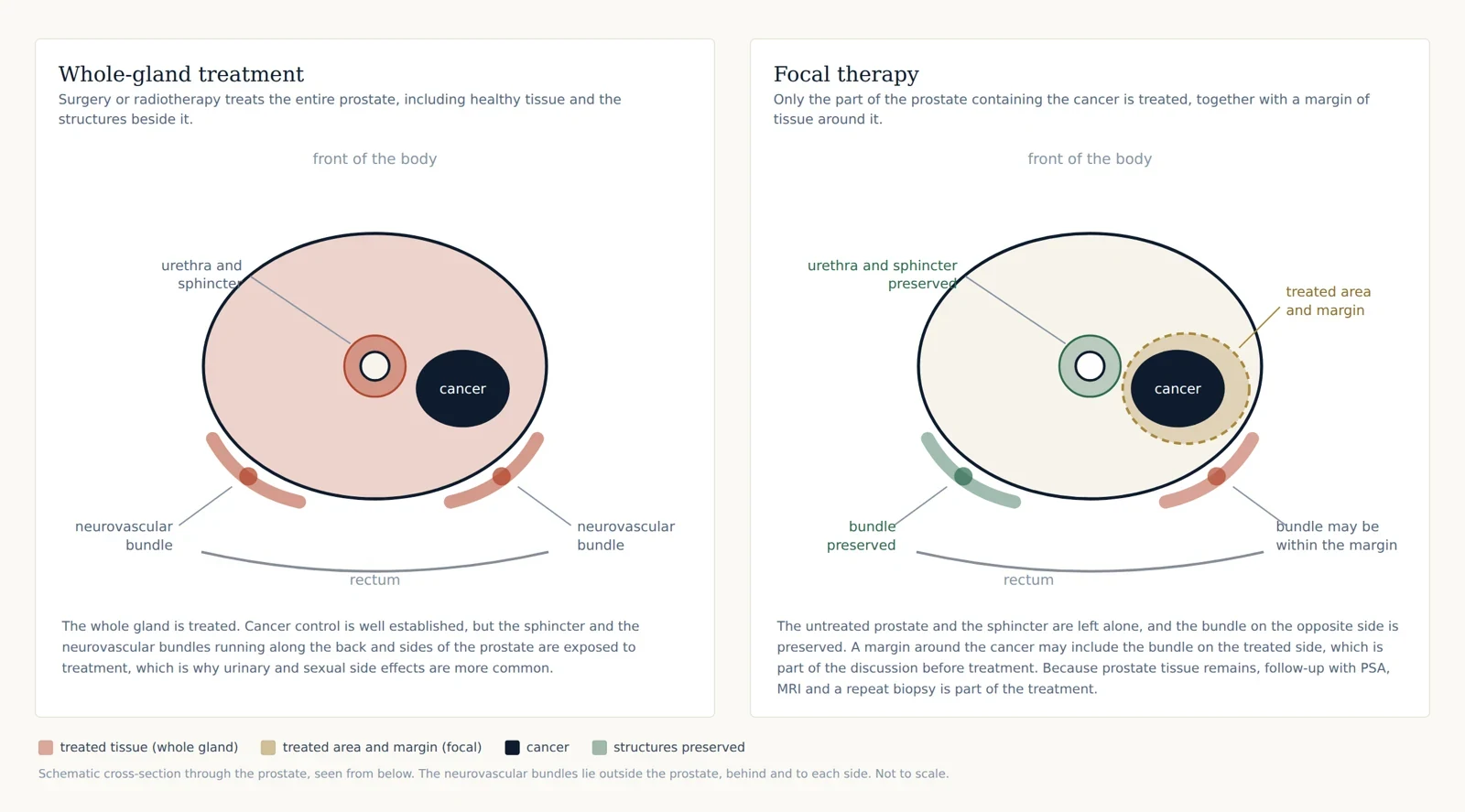
Prostate cancer has traditionally been treated by treating the whole prostate — removing it with surgery, or irradiating it. Both are effective but carry a risk of effects on urinary control and erections, because the whole gland and the structures around it are treated.
Focal therapy takes a different approach. Modern MRI and targeted biopsy can often show exactly where the significant cancer sits within the prostate. Where that area is well defined, it is possible to destroy only that part of the gland — together with a safety margin around it — and leave the rest of the prostate, the urinary sphincter and, where possible, the nerves responsible for erections untreated.
The tissue is destroyed in place rather than removed, using heat, cold or electrical pulses delivered through a probe or fine needles. No incision is made in the abdomen.
The aim is to control the cancer with fewer effects on daily life. The trade-off is that prostate tissue remains, so cancer can occasionally persist or appear later elsewhere in the gland, and lifelong monitoring is part of the treatment rather than an optional extra.
Where focal therapy currently sits
It is important to be clear about the evidence, because focal therapy is a developing field rather than a settled standard.
- The European Association of Urology recommends that focal ablation is offered within clinical trials or prospective registries.
- The American Urological Association and ASTRO state that whole-gland and focal ablation remain investigational, in the absence of high-quality trials comparing them directly with surgery, radiotherapy and active surveillance, and should not be used for high-risk disease outside a trial.
- NICE in the United Kingdom permits focal HIFU and irreversible electroporation under “special arrangements”, meaning careful patient selection, clear consent about the uncertainty, and prospective recording of outcomes. Focal cryotherapy is similarly covered by specific guidance.
- NCCN lists ablative techniques among the options for selected patients, again with the expectation of careful selection and follow-up.
In practice this means focal therapy is a reasonable option for carefully selected men who understand the uncertainty, want to avoid or defer whole-gland treatment, and accept a structured follow-up programme including repeat MRI and mandatory biopsy. It is not offered as an alternative to surgery or radiotherapy for men whose cancer is not suitable. Outcomes are recorded prospectively so that results can be reviewed.
Who it may suit
Suitability is decided from the MRI, the biopsy results, the PSA and PSA density, and your own priorities. Focal therapy is most likely to be appropriate when:
- the significant cancer is visible on MRI and confined to one clearly defined area, or one side of the prostate
- the grade or Gleason score is intermediate-risk disease — Grade Group 2 (Gleason 3+4), and selected Grade Group 3 (Gleason 4+3)
- biopsy confirms the cancer is where the MRI says it is, with no significant cancer elsewhere in the gland
- there is no evidence of spread beyond the prostate on MRI and, where indicated, PSMA-PET
- the size and shape of the prostate allow the target to be reached safely
- you are willing to attend regular PSA tests, repeat MRI and a planned repeat biopsy
Focal therapy is generally not suitable where the cancer is present on both sides in more than one significant area, where there is extensive high-grade disease, where the cancer has grown beyond the prostate, or where the whole gland is affected. In those situations, robotic prostatectomy or radiotherapy remain the appropriate treatments, and honest discussion about that is part of the assessment.
Low-risk disease (Grade Group 1) is usually best managed with active surveillance rather than treating it, focal or otherwise.
The energy sources used
Three energy sources have the most evidence behind them. The choice depends chiefly on where the cancer sits within the prostate, the shape and size of the gland, and whether the target lies close to the urethra, the rectum or the nerve bundles. HIFU and irreversible electroporation are the techniques used in this practice, and both are available at Subang Jaya Medical Centre.
High-intensity focused ultrasound (HIFU)
HIFU uses focused ultrasound waves to heat the target tissue to a temperature that destroys it, delivered through a probe placed in the back passage. Nothing passes through the skin.
The The HIFU system fuses your MRI images with live ultrasound, so the area to be treated is planned on the MRI and then treated under real-time ultrasound guidance. Treatment is delivered in small, controlled segments, and the ablation can be checked during and immediately after the procedure. HIFU is well suited to targets in the posterior part of the prostate, nearer the rectum, and its precision allows treatment close to structures that need to be preserved. Anterior targets can also be treated effectively in men with smaller prostates.
HIFU has the largest published experience of any focal technique, including medium-term multicentre outcomes.
Irreversible electroporation (IRE)
IRE is not a heat treatment. Fine needles are placed through the skin of the perineum, on either side of the target, and short, high-voltage electrical pulses are delivered between them. The pulses open permanent pores in the cell membranes, so the cancer cells die while the surrounding collagen framework — including nerves, blood vessels and the urethra — is largely preserved.
Because it is non-thermal and spares these structures, IRE is particularly useful for cancers sitting at the front of the prostate, close to the urethra, or immediately adjacent to the nerve bundles, where heat-based treatment would be harder to deliver safely.
The pulses are timed to the heartbeat, and full muscle relaxation is needed, which is why the procedure is done under general anaesthetic. Tell the team if you have a pacemaker, an implanted defibrillator or a heart rhythm problem, as this affects whether IRE is suitable and how it is planned.
Cryotherapy
Cryotherapy destroys tissue by freezing. Needles are passed through the perineum and an ice ball is created under ultrasound guidance, with warming catheters protecting the urethra. It has a long track record, and remains a reasonable option particularly for anterior tumours and for prostates that are less suited to HIFU.
Other techniques
Focal laser ablation, photodynamic therapy and microwave ablation have also been used. The published experience is smaller and the evidence weaker than for the three above.
Before your procedure
- Assessment: focal therapy is planned from a recent multiparametric MRI and from biopsy results, which are used to guide treatment. In some cases a prostate biopsy is repeated — for example where it was performed elsewhere — to be sure focal therapy is suitable for you.
- Discussion: all treatment options are discussed with you, and the alternatives — active surveillance, robotic prostatectomy and radiotherapy — are set out alongside focal therapy, so that the comparison is explicit.
- Fasting: fast for six hours before the procedure. Clear fluids are usually permitted up to two hours beforehand.
- Medicines: blood-thinning medication is paused as directed. Tell the team about all medicines, including supplements.
- Bowel preparation: for treatment delivered through the rectum, a small enema is given on the morning of the procedure.
- Antibiotics: a single dose of antibiotic is given at the time of the procedure.
- Getting home: you will have had a general anaesthetic, so arrange for someone to take you home and stay with you overnight.
On the day
Focal therapy is performed under general anaesthetic and takes around two hours, depending on the size and position of the area being treated. Most men go home the same day.
For HIFU, a probe is placed in the back passage, the planned treatment area is matched to your MRI, and the ablation is delivered in segments. For IRE and cryotherapy, fine needles are placed through the skin of the perineum under ultrasound guidance, and the treatment is delivered through them. In all three, a catheter is placed at the end of the procedure: the prostate swells after treatment, which would otherwise obstruct the flow of urine.
Risks and benefits
What focal therapy offers
What has to be weighed against it
Less long-term evidence. There is no completed randomised trial comparing focal therapy directly with surgery or radiotherapy, which is why guidelines describe it as investigational or restrict it to registries. In the largest UK series, 88% of men were free of metastatic disease and had not needed surgery or radiotherapy five years after treatment.
Cancer can persist or recur. Untreated prostate remains, so cancer can be found later in the treated area (in-field recurrence) or elsewhere in the gland (out-of-field recurrence). A proportion of men need repeat focal treatment, or go on to prostatectomy or radiotherapy. Surgery to remove the prostate after focal therapy can be more challenging, and carries a higher risk of rectal injury, incontinence and erectile dysfunction than surgery performed first. As with any treatment, there is a small risk of the cancer progressing to metastatic disease.
Follow-up is demanding. Regular PSA tests, repeat MRI and a planned repeat biopsy are part of the treatment, not optional additions.
Not suitable for everyone. If the assessment shows the cancer is more extensive than the MRI suggested, focal therapy will not be recommended.
Risks of the procedure itself
Aftercare and recovery
When to seek urgent advice
Contact the clinic, or go to the Emergency Department at Subang Jaya Medical Centre outside working hours, if you cannot pass urine after the catheter is removed, develop a fever or shivering, pass heavy bleeding or clots, or have worsening pain.
- Is my cancer suitable for focal therapy?
- Should I have focal therapy, or is active surveillance sufficient?
- Which energy source would you use in my case, and why that one?
- How does focal therapy compare with surgery and radiotherapy for my particular cancer?
- What is the chance I will need further treatment, and what would that involve?
- What follow-up will I need, and for how long?
- What happens if the biopsy at one year still shows cancer?