Understanding Upper Tract Urothelial Cancer
An uncommon urothelial cancer — the same family as bladder cancer — that begins in the lining of the kidney’s drainage system or the ureter.
A guide for patients: what upper tract urothelial cancer is, how it is found, and the main ways it is treated. It is an uncommon cancer, and understanding a few key terms early makes the conversations ahead easier.
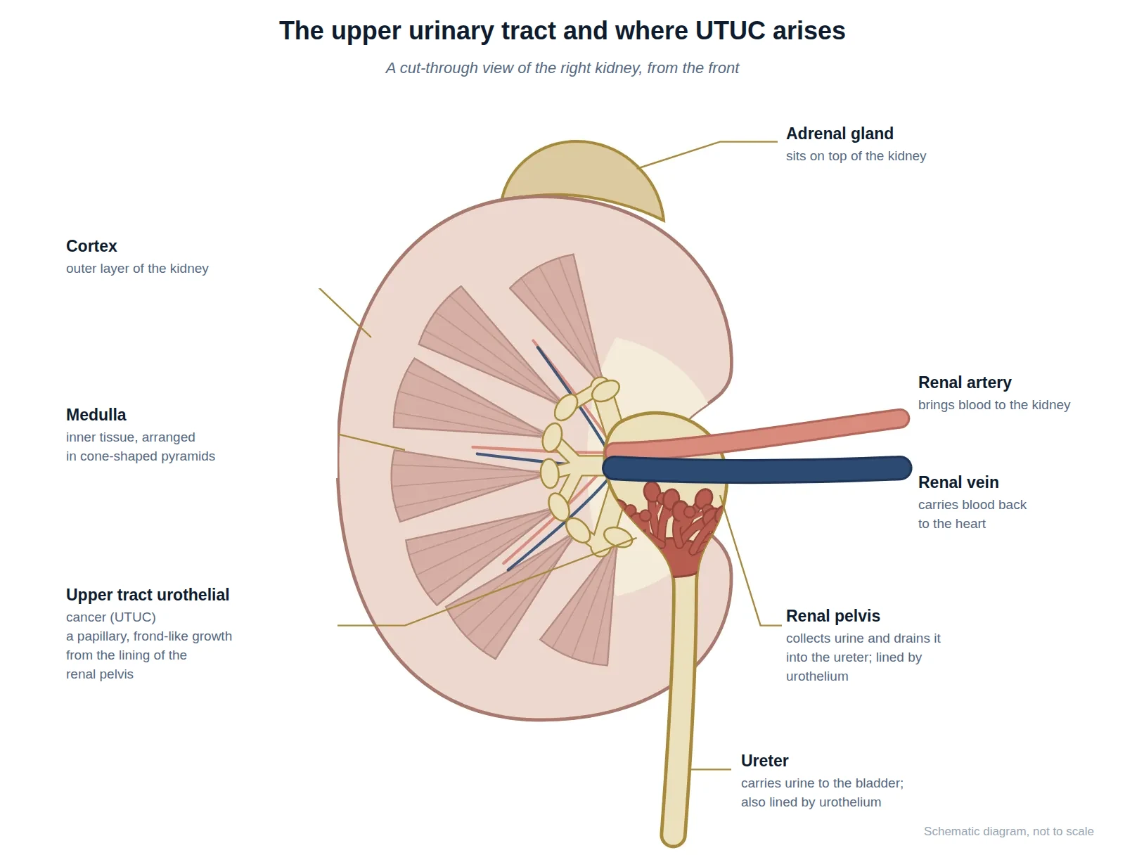
Urine made by the kidney collects in a funnel-shaped space called the renal pelvis, then travels down a thin tube — the ureter — to the bladder. These passages are lined by the same type of cells that line the bladder, called urothelium. When a cancer starts in this lining above the bladder, it is called upper tract urothelial cancer, or UTUC. This includes the renal pelvis and the ureter.
Because it is the same type of cell, UTUC is closely related to bladder cancer, and many of the same principles apply. It is much less common than bladder cancer, however, and its position deep inside the kidney and ureter makes it a little harder to diagnose and to treat.
Who it affects and why
UTUC becomes more common with age. The main risk factor is smoking. Previous bladder cancer also raises the risk, and a small number of cases are linked to inherited conditions (such as Lynch syndrome) or to certain historical exposures. Because UTUC and bladder cancer share the same lining, people treated for one are watched carefully for the other.
How it is found
The commonest first sign is blood in the urine. UTUC is usually identified with a CT scan of the urinary tract (a CT urogram), which outlines the drainage passages. To confirm the diagnosis and see the tumour directly, a thin flexible telescope is passed up into the ureter and kidney — a procedure called ureteroscopy — during which a small sample (biopsy) and washings can be taken. A urine sample may also be examined for abnormal cells.
Grade and stage
As with bladder cancer, two things guide treatment: the grade (how aggressive the cells look — low grade or high grade) and the stage (how deep the tumour has grown and whether it has spread). Low-grade, surface tumours behave gently; high-grade tumours, or those that have grown into the muscle wall of the ureter or kidney, are more serious and treated more aggressively.
Staging UTUC precisely can be difficult. The ureters are narrow, hence instruments used to visualise the ureter and take tissue samples are very small and may not reach deep enough to determine how far the tumour has invaded. Scans, too, are less precise. This means the exact stage is sometimes uncertain before removal of the kidney and ureter.
| Category | What it describes | Staging |
|---|---|---|
| T — Tumour | How deep the tumour has grown into the wall | Ta/CIS: urothelial lining T1: connective tissue invasion T2: into the muscle wall T3: through the wall into surrounding kidney/ureteric fat T4: grown into nearby organs or structures |
| N — Nodes | Whether it has reached nearby lymph glands | N0: no nodes involved N1–N2: lymph nodes involved |
| M — Metastasis | Whether it has spread to distant sites | M0: no distant spread M1: distant spread |
If it has spread
If UTUC has spread beyond the kidney and ureter, it is treated in the same way as urothelial bladder cancer that has spread, because they are the same kind of cancer. The first-line standard is a combination of newer drug treatments. You can read more on our page about metastatic bladder cancer.
Have a question about this condition?
Dr Tan consults at SJMC, Kuala Lumpur.
This cancer is often found after blood in the urine.