Kidney Stones
Why kidney stones form, how they are found and treated, and how to lower the chance of more stones.
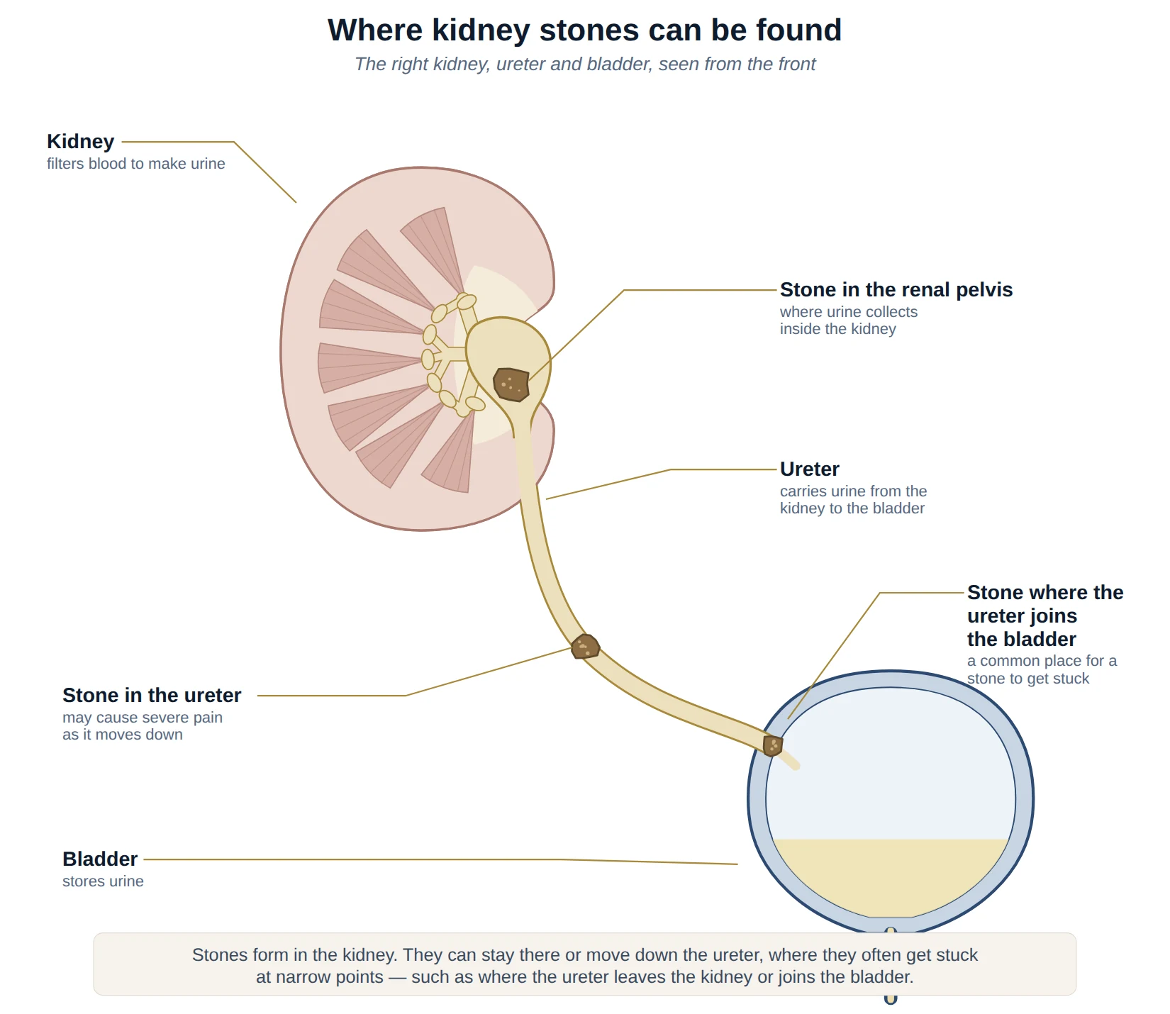
Kidney stones are hard deposits that form when minerals and salts in the urine crystallise and stick together. They range from the size of a grain of sand to several centimetres. Some stay quietly in the kidney and cause no symptoms; others move into the ureter — the tube that carries urine from the kidney to the bladder — where they can cause severe pain. Most small stones pass on their own, and for those that do not, there are effective treatments.
How common are kidney stones?
Kidney stones are common. In a large national survey in the United States, around 1 in 11 people had had a kidney stone — about 11% of men and 7% of women. Stones were more common in people living with obesity or diabetes. Stones are also more common in hot, tropical countries such as Malaysia, where dehydration is more likely.
Stones also tend to come back. In a study of more than 2,200 people followed after their first painful stone, 11% had another stone episode within 2 years and 20% within 5 years. This is why preventing new stones matters as much as treating the stone itself.
Symptoms
Stones that stay in the kidney often cause no symptoms and may be found by chance on a scan. When a stone moves into the ureter and blocks the flow of urine, it can cause:
- Renal colic: sudden, severe pain in the side or back, below the ribs, which often comes in waves and may spread to the lower abdomen or groin. It can be hard to find a comfortable position.
- Nausea and vomiting, often with the pain.
- Blood in the urine: visible or found only on a urine test — see blood in the urine.
- Urinary symptoms: needing to pass urine urgently or often, or discomfort, when a stone is close to the bladder.
Seek urgent medical help if you have stone pain together with a fever, shivering or feeling unwell, as this can mean an infection behind a blocked kidney, which needs emergency treatment. Also seek help promptly if you cannot control the pain or vomiting, are unable to pass urine, or have only one working kidney.
Types of stone and why they form
Stones form when the urine contains more stone-forming substances than it can keep dissolved — often because there is too little fluid, or too much of a particular substance. Knowing the type of stone helps guide prevention, so stones that are passed or removed are usually sent for analysis.
| Type of stone | How common | Common causes or links | Notes |
|---|---|---|---|
| Calcium stones (calcium oxalate or calcium phosphate) | Most common | Low fluid intake, a diet high in salt, and high levels of calcium or oxalate, or low levels of citrate, in the urine | Prevention focuses on fluids and diet; sometimes medication |
| Uric acid stones | Less common | Acidic urine; linked with gout, diabetes, obesity and a diet high in animal protein | Can often be dissolved with medication that makes the urine less acidic |
| Infection (struvite) stones | Less common | Certain urine infections | Can grow large and branch through the kidney (“staghorn” stones); usually need complete removal |
| Cystine stones | Rare | An inherited condition (cystinuria) | Often start at a younger age and tend to recur |
Risk factors
Several factors increase the chance of forming stones:
- Not drinking enough, and a hot climate: fluid lost through sweating makes the urine more concentrated. Living in a hot climate such as Malaysia increases the risk, so drinking enough is particularly important.
- Diet: a high intake of salt, animal protein and sugary drinks.
- Weight and metabolic health: obesity, diabetes and gout.
- A previous stone or a family history of stones.
- Certain medical conditions, such as an overactive parathyroid gland, some bowel conditions or previous bowel surgery, and some kidney conditions.
- Some medicines and supplements.
Investigations
Tests aim to confirm a stone, find its size and position, check for infection or a blockage, and look for reasons why stones are forming:
- Urine test: to look for blood and infection.
- Blood tests: including kidney function, calcium and uric acid.
- CT scan (low-dose, without contrast): the most accurate test for kidney stones, showing their size and position. It is usually the first scan when a stone is suspected.
- Ultrasound scan: avoids radiation, and is used for some patients and for some follow-up scans.
- X-ray: sometimes used to follow stones that show up clearly on X-ray.
- Stone analysis: identifying the type of stone passed or removed.
Metabolic assessment. For people at higher risk of forming more stones — for example, those with recurrent stones, stones at a young age, a strong family history or certain stone types — a more detailed assessment may be recommended, including a 24-hour urine collection to measure the substances that cause stones.
Will the stone pass on its own?
Many small stones pass without treatment, usually within a few weeks. The chance depends mainly on the size of the stone and where it is: smaller stones, and those lower down the ureter nearer the bladder, are more likely to pass.
| Stone size or position | Passed without a procedure |
|---|---|
| By stone size | |
| Under 5 mm | About 89% |
| 5–7 mm | About 49% |
| Over 7 mm | About 29% |
| By position in the ureter | |
| Lower ureter (nearest the bladder) | About 83% |
| Middle ureter | About 70% |
| Upper ureter (nearest the kidney) | About 52% |
Source: the MIMIC study of 2,518 patients with a single ureteric stone confirmed on CT, initially managed without a procedure, across 71 hospitals in the UK, Ireland, Australia and New Zealand (Shah et al., BJU International, 2019). “Passed without a procedure” means the stone did not need an operation to remove it.
While waiting for a stone to pass, pain is usually controlled with anti-inflammatory painkillers, and you may be asked to strain your urine to catch the stone for analysis. A follow-up scan checks whether it has passed. If it has not passed, if pain is uncontrolled, or if there are signs of infection or reduced kidney function, treatment is recommended.
Medicine to help a stone pass. Tamsulosin, which relaxes the muscle of the ureter, is sometimes prescribed to help a stone pass. A large UK randomised trial (SUSPEND, more than 1,100 patients) found no overall benefit compared with a placebo. However, a large randomised trial of 3,296 patients in China found that tamsulosin increased the proportion of stones in the lower ureter that passed (86% compared with 79%), with the benefit seen in stones larger than 5 mm, and also reduced pain. Because tamsulosin is a low-risk medication, it is still widely used for stones in the lower ureter.
Stones that cause no symptoms
Small kidney stones are often found by chance on a scan done for another reason. Many never cause problems, and these can usually be monitored with occasional scans rather than treated straight away.
Treatment may be recommended if a stone:
- grows, or starts to cause pain, infection or blood in the urine
- blocks the flow of urine from the kidney
- is large, or is in a single working kidney
- would be a particular problem if it caused sudden pain — for example, for pilots, people who travel frequently or work far from medical care
The decision balances the chance of future problems against the small risks of treatment, and your own preferences.
Treatment options
Treatment depends on the size, position and type of stone, whether it is causing symptoms or a blockage, and your preferences.
| Treatment | When it is used |
|---|---|
| Watchful waiting | Small stones likely to pass on their own, or small kidney stones causing no symptoms |
| Tamsulosin | To help stones in the lower ureter pass |
| Emergency drainage (stent or nephrostomy) | A blocked kidney with infection, or other urgent situations; the stone is treated once the infection has settled |
| Shockwave lithotripsy (ESWL) | Stones under 2 cm in the kidney or upper ureter |
| Ureteroscopy with laser (URS) | Stones in the ureter, and small to medium kidney stones |
| Percutaneous nephrolithotomy (PCNL) | Large or complex kidney stones, typically over 2 cm |
| Dissolution therapy | Uric acid stones, using medication that makes the urine less acidic |
Each linked treatment opens its own section on the Kidney Stone Procedures page, with what it involves, its risks and recovery.
Ureteric stents
A ureteric stent is a thin, soft, hollow tube placed inside the ureter, with one end curled in the kidney and the other in the bladder. It keeps urine draining from the kidney and is used when a stone is causing significant blockage of the kidney. A stent is often left in for a short time after ureteroscopy to allow swelling to settle.
What to expect. Stents are not usually painful, but many people notice them. Common symptoms include needing to pass urine more often or urgently, discomfort in the bladder or in the side — particularly when passing urine — and blood in the urine. These symptoms settle once the stent is removed.
Removal. Most stents are removed after a short period, usually with a quick flexible cystoscopy in the clinic, or sometimes by gently pulling a thread left attached to the stent. It is important that a stent is removed or changed on time: a stent left in too long can become coated with crystals and block, or be difficult to remove.
Please contact the team if you develop a fever, severe pain or heavy bleeding with a stent in place.
Preventing further stones
Lifestyle changes can substantially lower the chance of forming new stones:
- Drink plenty of fluid: enough to pass at least 2.5 litres of urine a day. For most people this means drinking around 2.5 to 3 litres a day, spread through the day — and more in hot weather or when exercising. Pale urine is a helpful guide. Water is best.
- Reduce salt: too much salt increases the calcium in the urine. Limit salty and processed foods.
- Keep a normal calcium intake: around 1,000–1,200 mg a day, ideally from food such as dairy. Do not cut out calcium unless advised, as too little calcium in the diet increases the absorption of oxalate and the risk of calcium oxalate stones.
- Moderate animal protein: such as red meat, poultry and seafood.
- Eat plenty of fruit and vegetables: these increase citrate in the urine, which helps prevent stones. Adding lemon or lime juice to water may also help.
- If you have calcium oxalate stones, limit foods very high in oxalate — such as spinach, rhubarb, beetroot, nuts, chocolate and strong tea — and have calcium-containing foods with meals.
- Limit sugary and fizzy drinks, and keep to a healthy weight.
Medicines to prevent stones. For people who continue to form stones despite these measures, medication may be recommended based on the stone type and urine tests:
- Thiazide medicines: lower the amount of calcium in the urine.
- Potassium citrate: raises citrate in the urine and makes it less acidic — used for low urine citrate, and for uric acid and cystine stones.
- Allopurinol: lowers uric acid, for people with high levels of uric acid who form calcium oxalate stones.
Have a question about this condition?
Dr Tan consults at SJMC, Kuala Lumpur.